3.1 The Evolution of Bodies and Brains
Brains did not appear on their own and then acquire bodies. They evolved within bodies that could already hold themselves together and act in their surroundings. Following that history is important for understanding development because nervous systems and brains emerged through changes in the developmental organization of whole animals, not as structures added to otherwise finished bodies.
Although we often think about the brain as the control center for the body, bodies existed well before brains. Multicellular bodies came first, neurons developed later as specialized cells within them, and brains appeared later still. At each point, evolutionary change occurred through changes in development, as existing cellular capacities were elaborated and reorganized within new kinds of bodies. New capabilities emerged, but they were built from processes and forms of organization with much older histories.
The sequence that follows is not a ladder leading toward the vertebrate or human brain. The question is what had to change in the development and organization of animal bodies before a brain could become part of them. Following these changes gives a clearer sense of what nervous systems and brains evolved to do, and it turns out to challenge some surprisingly basic assumptions about what brains are for.
The Origins of Neurons
Before there were neurons and nervous systems, before there were bodies, and even before there were animals, much of the molecular machinery that neurons rely on already existed. Voltage-gated ion channels and other components later used in neural signaling have evolutionary roots that predate animal nervous systems (Liebeskind et al., 2011, 2017). Molecules now used in communication across synapses likewise have deeper histories in the regulation of cellular activity, the coordination of neighboring cells, and the maintenance of internal organization.
Evolution did not invent an entirely new chemistry when neurons appeared. What was already there was repurposed and elaborated. A neuron is a cell exquisitely specialized for rapid, directed signaling, but the capacities on which that specialization depends are much older. Living cells could already respond to changes around them and communicate chemically, long before anything resembling a nervous system appeared.
So why did neurons evolve at all? A single-celled organism has no coordination problem: it is already a unity. A large multicellular body, made of cells that have taken on different functions in different places, has to find a way to act as a whole. As animal bodies became larger and more differentiated, communication across the body mattered more and more. Neurons offered a way for activity in one region to alter activity elsewhere rapidly and selectively.
Nervous systems are often described in input-output terms: something happens outside the animal, the nervous system processes information about it, and the animal responds. But sensitivity to surrounding conditions did not begin with neurons. One family of proposals is that nervous systems first became important within the problem of coordinating a multicellular body (Arendt, 2021; Fields et al., 2020). This does not mean that early nervous systems were purely internal and acquired sensory functions only later. Sensing, movement, and bodily coordination probably developed together. The point is that a nervous system belonged to a body before it could become a system for representing a world outside that body.
Jellyfish show what a nervous system can look like without a brain. Their nervous systems include nerve nets and, depending on the group, nerve rings or localized sensory and coordinating centers. These systems coordinate swimming and other activities without a single central brain (Cunningham et al., 2024). Neurons, then, do not by themselves imply a brain. Jellyfish occupy a different region of the space of animal possibilities, one that has supported a remarkably durable way of life. We will return to them at the end of the reading.
Bilateral Bodies and a Leading End
Living animals are commonly organized into five major clades: Porifera, which includes sponges; Ctenophora, the comb jellies; Placozoa; Cnidaria, which includes jellyfish, sea anemones, and corals; and Bilateria. Humans belong to Bilateria, along with the great majority of the animals that are most familiar to us. When we think of animals, we tend to think of bilaterians, which can make their organization seem like the definition of an animal body. It is only one of several enduring ways of being an animal.
As their name suggests, bilaterians are organized around bilateral symmetry. An anterior-posterior axis runs from front to rear, while a plane of symmetry divides the body into corresponding left and right sides. Other animals may instead be organized radially around an axis, as in jellyfish, or lack a fixed symmetry, as in many sponges. These arrangements do not prevent directional movement. In many bilaterian lineages, however, sustained movement became organized around a relatively stable leading end. That end of a moving animal was usually the first part to meet food, danger, or anything else that mattered. This opened new possibilities for coordinating movement and sensory activity, and for concentrating sensory structures and neural tissue toward the anterior end.
Bilaterians were present by the late Ediacaran, more than 550 million years ago, and subsequently diversified into an extraordinary range of forms (Erwin et al., 2011). What made that diversification possible? There is no single answer, but part of it lies in changes to the developmental processes through which the internal organization of animal bodies was built. To see why, we need to look at something that happens early in embryonic development: gastrulation.
Gastrulation and the Germ Layers
An embryo does not begin with its tissues and organs already in place. During gastrulation, cells move. Some move toward the interior of the embryo, while others spread across its surface. As they move, they come into contact with new neighbors and become exposed to different signals. The details vary across animals, but in bilaterians these movements establish three broad populations of embryonic cells known as the germ layers: ectoderm, endoderm, and mesoderm. They are not permanent layers of the adult body but changing populations of cells that contribute to different tissues and organs as development continues. Having three germ layers makes bilaterians triploblastic. Cnidarians such as jellyfish are usually described as diploblastic, with two.
Broadly speaking, ectoderm contributes to the outer covering of the body and the nervous system, while endoderm contributes to the digestive tract and associated organs. Mesoderm contributes to muscle, connective and supporting tissues, circulatory structures, and many internal organs. Mesoderm was not a new kind of biological material. The cells that formed it relied on older capacities for movement, adhesion, signaling, and differentiation; what was new was how those capacities worked together in development to produce, reliably, a population of cells with new relations to the rest of the embryo.
Why is this important for the evolution of bodies and brains? The organization of mesoderm opened new possibilities for more extensive musculature and internal organization, while ectoderm gave rise to the nervous system in relation to that changing body. This did not prescribe the appearance of a worm, an insect, an octopus, or a vertebrate. It changed the developmental starting point from which those very different bodies could arise. Like the neuron, the organization of the germ layers was new, but it was made from cellular activities that already had a history.
The Evolution of Brains
With multicellular bodies established and neurons in place, we can now ask how brains emerged from that foundation. The nerve net of a jellyfish shows that a body can coordinate movement and respond to its surroundings without concentrating neurons into a brain. Brains became possible as bodily coordination became increasingly intertwined with sensory activity, especially in animals whose movement repeatedly brought one part of the body into contact with what lay ahead.
In many bilaterian lineages, the leading end became a recurring location for coordinating feeding, sensory activity, and movement. Neural tissue and sensory structures became increasingly concentrated there. This is cephalization. The word can make it sound like a simple shift of neurons toward the front, but it was part of the development of a head: an organized anterior region in which sensory structures, feeding structures, muscles, supporting tissues, and neural activity came into new relations.
Complex brains appear to have evolved independently in vertebrates, arthropods, and the cephalopod branch of the mollusks (Moroz, 2009). A fish, an insect, and an octopus did not inherit different versions of one complex brain from a common ancestor. Each lineage developed its own way of coordinating an active, sensing body. A brain is therefore not a single evolutionary solution that some animals possess in a more advanced form than others.
There is a larger point here about what brains are for. A brain belongs to an animal that is already moving and acting, and that activity partly determines what its sensory systems encounter. The standard picture starts with information arriving from an external world and treats the brain as the processor that decides what the body should do. The evolutionary history suggests that this picture has things backwards. Brains evolved in animals that were already moving and acting, and the input a moving animal receives is partly a product of its own movement.
The relation between brain and body is reconstructed in every generation. Changes in the development and use of a body alter the conditions under which its nervous system develops, while changes in neural organization alter what the animal can do. This is why the evolution of bodies and the evolution of brains belong to the same story. Both concern changes in developmental processes that produce active organisms with particular ways of living.
The Cambrian and the Chordate Lineage
Cephalization followed different paths in vertebrates, arthropods, and cephalopods. We follow the chordates here. This is not because the vertebrate path is the best one, but because it is our path, and tracing it takes us from small suspension feeders in Cambrian seas to the brain you are using to read this sentence.
The Cambrian period, roughly 540 to 485 million years ago, saw a striking diversification of animal forms. It is often described as the Cambrian Explosion, although the term can be misleading. Animals, including bilaterians, were already present during the preceding Ediacaran period, and the developmental processes that made Cambrian bodies possible had deeper histories. What becomes especially visible in the Cambrian fossil record is a much wider range of recognizable animal organizations, together with shells, plates, and other hard structures that were more readily preserved (Erwin et al., 2011).
Arthropods, mollusks, worms, echinoderms, and early chordates were among the forms that diversified during this period. Their bodies were organized around different arrangements of segments, appendages, muscles, feeding structures, and sensory systems. Their anatomical differences changed what they could do.
The conditions in which this happened were not constant. Oxygen availability and ocean chemistry varied across time and place (Pruss & Gill, 2024), while changes in sea level altered the extent and character of habitable waters. No single environmental change explains the Cambrian diversification. These conditions helped shape what forms of metabolism, body size, and movement could be sustained, but animals also changed the conditions encountered by other organisms through feeding, movement, burrowing, competition, and predation.
Among this diversity were the early chordates. The ancestors of vertebrates were probably small, soft-bodied animals that lived in shallow water and filtered suspended nutrients. They would not have stood out beside the more conspicuous arthropods and other animals of Cambrian seas. Nobody looking at them then would have picked them out as the lineage that would eventually produce lampreys, frogs, pigeons, and people, and their organization did not contain that future waiting to emerge.
What distinguished chordates was the relation among a flexible supporting notochord, a dorsal nerve cord, segmented musculature, and the rest of the body. This organization supported swimming, with neural activity coordinating movements in response to surrounding conditions. The later emergence of vertebrates involved a transformation of this chordate organization, especially at its anterior end. The history of the vertebrate brain lies within that transformation of the body as a whole.
Brains Are for Moving Animals
A living relative of vertebrates, the sea squirt, provides one of the clearest illustrations of the relation between nervous systems and ways of life. Sea squirts such as Ciona begin life as small, free-swimming larvae with a simple brain and nerve cord. A larva moves through the water, orients itself, and eventually finds a surface to which it can attach. It then transforms into a sedentary adult filter feeder. Horie and colleagues (2011) showed that the tail nerve cord is lost and most larval neurons disappear, while other cells of the larval central nervous system remain and help build the adult nervous system. There is a familiar joke among biologists that the sea squirt eats its own brain when it settles down, but the actual change involves reconstruction as well as loss.
The adult keeps a nervous system suited to a sedentary life, but the larval brain that coordinated swimming is largely gone. Neural tissue is expensive to maintain, and an animal that has stopped moving no longer needs much of what that brain did. Brains and movement go together. As an animal moves, what it senses changes, and its next movements depend on those changes.
Stronger swimming, more differentiated sensory organs, and more neural tissue at the anterior end can look like separate additions, but they did not evolve independently and then get assembled together. A change in movement altered what the animal encountered. Changes in sensory activity altered the conditions under which neural organization developed. New forms of neural coordination changed what the animal could do.
From Chordates to the Vertebrate Head
Amphioxus, also known as the lancelet, is a living chordate that helps us think about the organization from which vertebrates evolved. It is not a surviving vertebrate ancestor or a relic unchanged since the Cambrian. It belongs to a separate lineage with its own long evolutionary history. This small, blade-shaped animal lives in coastal waters, where it burrows into sand and filters nutrients from the water.
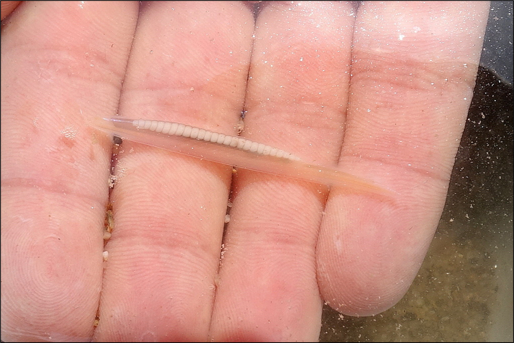
Photo © Pascal GIRARD, via iNaturalist (CC BY-NC 4.0)
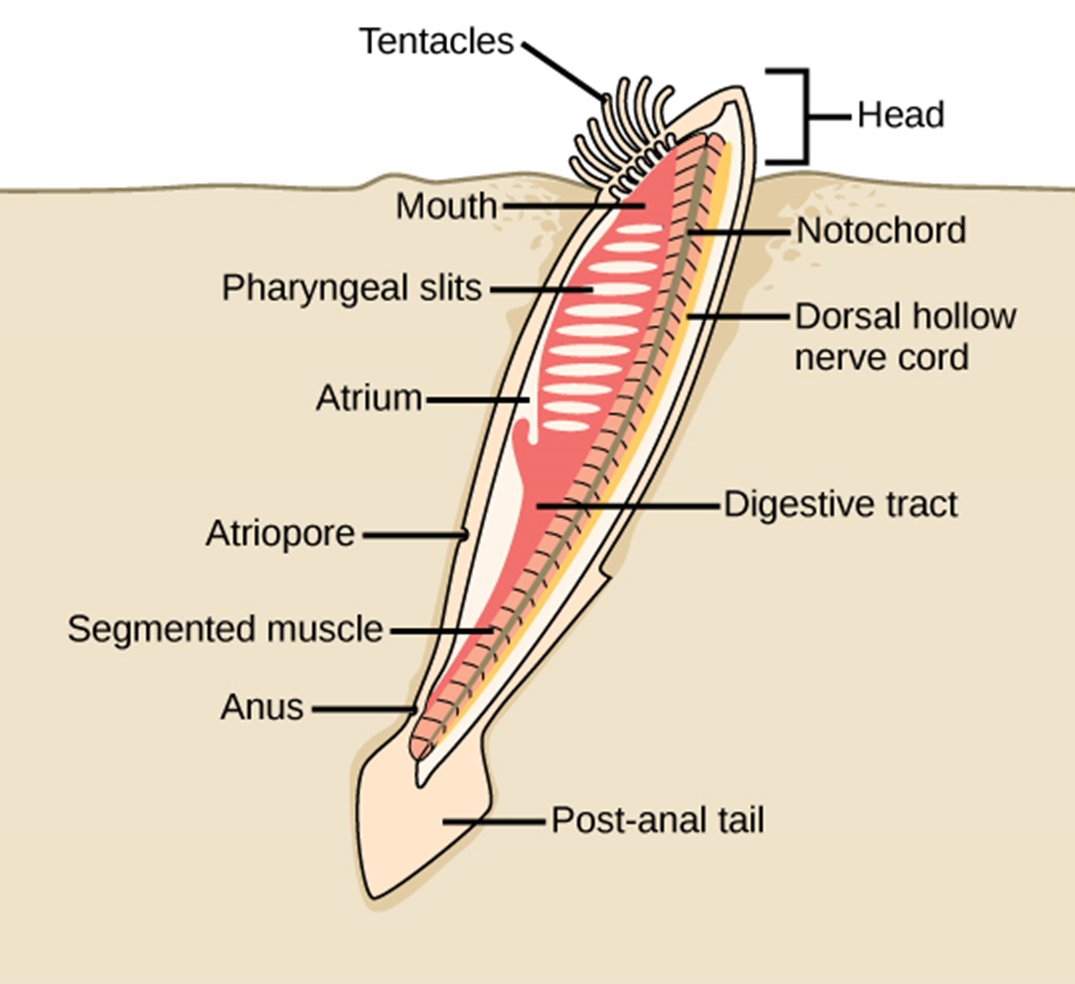
OpenStax, Concepts of Biology, Figure 15.35, via Biology LibreTexts (CC BY 4.0).
Amphioxus has a notochord, a dorsal nerve cord, and segmented muscle blocks that support swimming, while the anterior end of its nerve cord is only modestly enlarged. It has light-sensitive structures but lacks the image-forming eyes, vestibular system, and elaborate olfactory structures found in vertebrates. Amphioxus therefore shows us a chordate organization in which a notochord and dorsal nerve cord are present but the anterior body has not acquired the organization of a vertebrate head.
So how did things go from an ancestral chordate organization resembling Amphioxus in some respects to the vertebrate head? The front of the neural tube expanded, sensory structures developed around it, and muscles, nerves, and supporting tissues became organized within a distinct anterior region. These changes occurred together. The vertebrate brain emerged through this reorganization of the head and body.
An important part of this transformation involved neural crest cells. These cells form near the edges of the developing neural plate and then migrate through the embryo. In the head, their descendants contribute to the bones and cartilage of the face and skull, as well as to parts of the cranial and peripheral nervous systems. The cellular and molecular capacities involved had deeper roots among chordates, but their organization in vertebrate development helped make a head possible (Martik & Bronner, 2021). Neural crest contributes so much to the vertebrate head that it is sometimes called the fourth germ layer. It is also another case of something new emerging from a reorganization of capacities with older histories.
The Radiation of Fishes
The evolutionary path from the earliest vertebrates to mammals, birds, reptiles, and amphibians runs through fishes. The vertebrate head and brain first diversified in animals living in water. Some early vertebrates lacked jaws and paired fins, but they already possessed a more differentiated anterior body than other chordates. The brain, cranial sensory structures, pharyngeal region, and swimming musculature developed as parts of the organization through which these animals moved, fed, and oriented themselves.
The later emergence of jaws transformed this organization further. The jaw skeleton develops from the first, or mandibular, pharyngeal arch. Shared developmental features of this arch and the gill-bearing arches support the proposal that jaws evolved through changes to an ancestral gill arch (Thiruppathy et al., 2022). New ways of feeding developed together with changes in movement, sensory activity, and neural coordination. Jawed vertebrates subsequently radiated into the lineages that became cartilaginous and bony fishes. Some of this history is still in your own head: the small bones of the mammalian middle ear derive from elements of the jaw joint of our early mammalian ancestors (Shubin, 2008).
This radiation also transformed sensory life. Image-forming eyes, increasingly differentiated vestibular and olfactory systems, and forms of mechanical and electrical sensing gave fishes access to different features of the surrounding water. The aquatic environment was not a single setting. Differences in depth, light, current, substrate, and the activity of other organisms created different conditions for moving, feeding, and orienting. Sensory systems and brains developed within these different ways of living rather than as improvements added one at a time.
The history of fishes was not a progression toward life on land. Fishes diversified into many enduring forms, most of which had nothing to do with the later emergence of tetrapods. One branch of lobe-finned fishes eventually gave rise to animals with limbs capable of supporting movement in shallow water and on land. Apart from those tetrapod descendants, lobe-finned fishes survive today only as a handful of lungfish species and the coelacanth, a deep-sea fish thought to be extinct until one was caught off South Africa in 1938. Amphibians, reptiles, birds, and mammals therefore emerged from within a much older vertebrate history in water. Our heads and brains still bear that history, even though the inherited organization has been modified through very different bodies and ways of life.
Epilogue: A Meditation on the Jellyfish
In following a path from multicellular bodies to neurons, bilateral organization, cephalization, and vertebrate brains, it is easy to see animals without brains as having been left behind. The sequence can look like a history of progress, as though complexity were the destination and everything else represented a failure to arrive. But evolution has no destination. Each change in development altered what was possible, and what emerged was one path through that space rather than the goal toward which animal life had been moving.
Jellyfish and bilaterians have been evolving for approximately the same length of time. Jellyfish have continued to thrive without a centralized brain, through an organization that coordinates movement and activity across a very different kind of body. They have persisted through changing oceans and repeated mass extinctions. The jellyfish pulsing through the ocean is not a lesser form of animal, and its way of life rests on a relation between body and world that has stayed viable for hundreds of millions of years.
The diversity of life includes many forms that have flourished without anything resembling a vertebrate brain. Complexity is one path through the space of possibilities, but it is not the only one and may not be the most durable. The jellyfish is a reminder that there is more than one way to persist within it. And for jellyfish, it has worked pretty well.
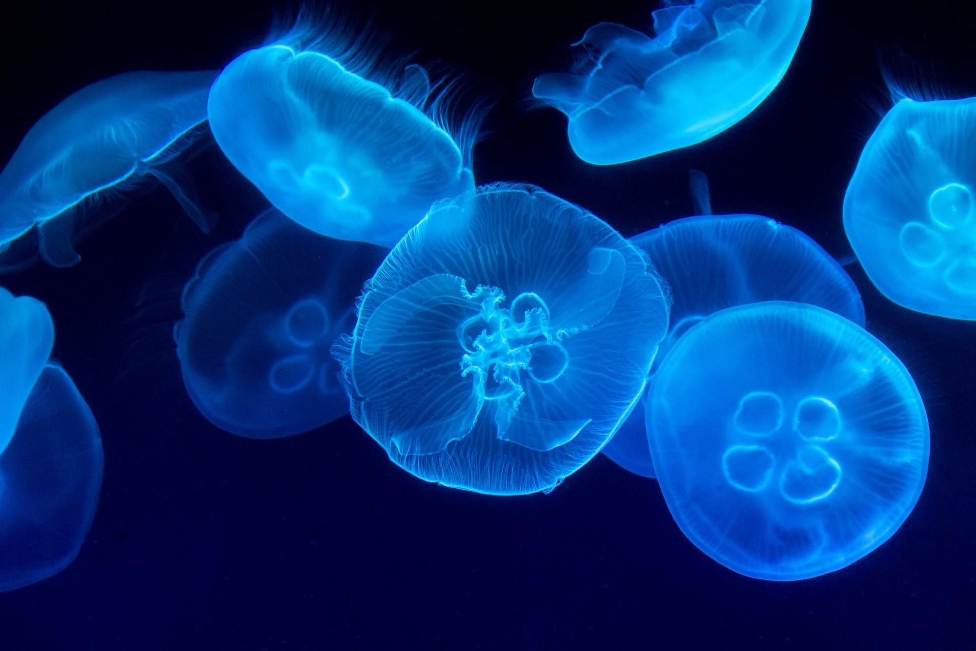
Photo by Marat Gilyadzinov on Unsplash. Used under the Unsplash License; Excluded from this resource’s CC BY-NC-SA 4.0 license.
Sources
Arendt, D. (2021). Elementary nervous systems. Philosophical Transactions of the Royal Society B: Biological Sciences, 376(1821), 20200347. https://doi.org/10.1098/rstb.2020.0347
Cunningham, K., Anderson, D. J., & Weissbourd, B. (2024). Jellyfish for the study of nervous system evolution and function. Current Opinion in Neurobiology, 88, 102903. https://doi.org/10.1016/j.conb.2024.102903
Erwin, D. H., Laflamme, M., Tweedt, S. M., Sperling, E. A., Pisani, D., & Peterson, K. J. (2011). The Cambrian conundrum: Early divergence and later ecological success in the early history of animals. Science, 334(6059), 1091–1097. https://doi.org/10.1126/science.1206375
Fields, C., Bischof, J., & Levin, M. (2020). Morphological coordination: A common ancestral function unifying neural and non-neural signaling. Physiology, 35(1), 16–30. https://doi.org/10.1152/physiol.00027.2019
Horie, T., Shinki, R., Ogura, Y., Kusakabe, T. G., Satoh, N., & Sasakura, Y. (2011). Ependymal cells of chordate larvae are stem-like cells that form the adult nervous system. Nature, 469, 525–528. https://doi.org/10.1038/nature09631
Liebeskind, B. J., Hillis, D. M., & Zakon, H. H. (2011). Evolution of sodium channels predates the origin of nervous systems in animals. Proceedings of the National Academy of Sciences, 108(22), 9154–9159. https://doi.org/10.1073/pnas.1106363108
Liebeskind, B. J., Hofmann, H. A., Hillis, D. M., & Zakon, H. H. (2017). Evolution of animal neural systems. Annual Review of Ecology, Evolution, and Systematics, 48, 377–398. https://doi.org/10.1146/annurev-ecolsys-110316-023048
Martik, M. L., & Bronner, M. E. (2021). Riding the crest to get a head: Neural crest evolution in vertebrates. Nature Reviews Neuroscience, 22, 616–626. https://doi.org/10.1038/s41583-021-00503-2
Moroz, L. L. (2009). On the independent origins of complex brains and neurons. Brain, Behavior and Evolution, 74(3), 177–190. https://doi.org/10.1159/000258665
Pruss, S. B., & Gill, B. C. (2024). Life on the edge: The Cambrian marine realm and oxygenation. Annual Review of Earth and Planetary Sciences, 52, 109–132. https://doi.org/10.1146/annurev-earth-031621-070316
Shubin, N. (2008). Your inner fish: A journey into the 3.5-billion-year history of the human body. Pantheon Books.
Thiruppathy, M., Fabian, P., Gillis, J. A., & Crump, J. G. (2022). Gill developmental program in the teleost mandibular arch. eLife, 11, e78170. https://doi.org/10.7554/eLife.78170