3.2 Brain, Body, World
The vertebrate brain has a shared developmental organization whose evolutionary history extends deep into the past. Here we change scale and ask how that organization is reconstructed in a single developing individual. The focus is the cortex, where the relation between reliable organization and developmental activity can be seen especially clearly. The way cortical organization takes shape also makes it difficult to treat brain, body, and surrounding world as separate sources.
From Fertilization to the Neural Tube
Human embryonic development begins with fertilization, forming a single cell (the zygote) that then undergoes cleavage. By day 3, the embryo reaches the morula stage (16 to 32 cells). Around day 4 to 5, it has developed into a blastocyst (50 to 150 cells), with two distinct cell populations: the outer trophoblast and the inner cell mass. Implantation in humans begins around day 6 to 7 after fertilization, as the blastocyst attaches to the uterine wall, a process that takes several days to complete (Lumen Learning, n.d.).
Gastrulation typically begins around day 14 to 16 after fertilization. It is a critical process that reorganizes the embryo and lays the foundation for the body and brain. During gastrulation, cells from the epiblast, which is derived from the inner cell mass, migrate to form the primitive streak. The primitive streak is an early landmark of the embryo’s body plan and anterior-posterior organization. As gastrulation proceeds, cell migration through the primitive streak establishes endoderm and mesoderm, while epiblast cells remaining at the surface form ectoderm.
During gastrulation and neurulation in vertebrates, signals exchanged among embryonic tissues help establish the neural plate. The notochord, a mesodermal structure beneath it, contributes to the patterning of the developing nervous system. The neural plate folds to form the neural tube, the embryonic structure that becomes the central nervous system. In humans, the main process of neural tube closure is complete around 4 weeks after fertilization. The anterior region becomes the brain, while more posterior regions become the spinal cord (Darnell & Gilbert, 2017).
As the neural tube develops, neural progenitor cells multiply in the ventricular zone lining its cavity. In the developing cortex, additional progenitors divide in the subventricular zone. Early divisions expand the progenitor population; later divisions also produce neurons, directly or through intermediate progenitors. Newly formed neurons generally stop dividing before migrating toward their eventual positions, while other progenitors later produce glia. As in every other part of the body, what a cell born into the developing nervous system becomes depends on when and where it was produced within the embryo, and on the signals it receives from neighboring cells.
Side Note: Insights from Embryology
Not about the brain specifically, but some of the classic research on how the “when and where” influences the development of embryonic cells came from Hans Spemann and his collaborators in the early twentieth century. Spemann showed that tissue exchanged between early newt gastrulae could develop in accordance with its new location, whereas tissue transplanted at later stages was more likely to retain the fate it had already been taking on (Spemann, 1935).
Related work by Hilde Mangold with Spemann showed that a transplanted region from the dorsal lip of the blastopore could induce surrounding host tissue to form a secondary body axis, establishing the concept of an embryonic “organizer” (Spemann & Mangold, 1924/2001). The transplantation experiments tell us something about a window for plasticity in what developing cells become. The organizer experiment also shows how one region changes the conditions for surrounding tissue. We will return to organizers at the end of the reading, when we ask where the organizer itself comes from.
The timing and process of gastrulation vary across animals, reflecting different reproductive strategies and types of life cycle. In humans and other mammals, some of what unfolds during early embryonic development reflects adaptations related to the placenta and to live birth. Even so, we can learn a great deal by looking at basic developmental processes in other organisms.
Building the Neocortex
During neurulation, the anterior portion of the neural tube expands to form three primary brain vesicles: the prosencephalon (forebrain), mesencephalon (midbrain), and rhombencephalon (hindbrain). The prosencephalon further divides into the telencephalon and diencephalon. The telencephalon gives rise to the cerebral hemispheres, including the neocortex. We focus here on the neocortex because its development raises some of the central questions about brain development more generally, including how neural activity contributes to its organization.
In humans, around 90% of the cortex is neocortex, with the other 10% being phylogenetically older allocortex (mainly olfactory cortex and the hippocampus). The human neocortex is a sheet of tissue a few millimeters thick and around 2,400 square centimeters in area, folded into the indentations (sulci) and ridges (gyri) that give the brain its characteristic appearance. It has a layered structure, being composed of six layers that each have a particular configuration of neurons, glia, and connectivity.
Many excitatory neurons of the neocortex are produced in ventricular and subventricular zones and migrate outward toward the cortical plate. Most cortical layers are built from the inside out: earlier-born neurons form deeper layers, and later-born neurons migrate past them to form more superficial layers. Most human neocortical neurons are produced before birth (Jernigan & Stiles, 2017). Once situated in a layer, adhesion molecules help neurons stay together in functional units that are a product of their location in the cortex, the layer they belong to, and their connectivity with other neurons across the cortex and in subcortical areas.
In most mammals, migrating neurons pass through a subplate on their way to the cortical layers. In humans, the subplate expands during fetal development and later diminishes as cortical connections are reorganized; some of its neurons persist into adulthood (Kostović & Rakic, 1990). Its role is still being studied, but subplate neurons participate in early electrical activity and synaptic connections.
Neurons are generating electrical activity and forming functional circuits while the cortex is still being assembled. The brain is not built first and switched on later: it is active while it is being built, and that activity influences how it develops.
Cortical Areas and the Bodies They Reflect
As well as having a layered structure, the neocortex is composed of many regions that vary in their microstructural and functional properties. The most obvious are the primary sensory areas, such as primary visual cortex (V1), primary auditory cortex (A1), and primary somatosensory cortex (S1), but there are many more. How many depends on the level at which you are looking. The classic scheme of Brodmann identifies 52 areas based on cytoarchitecture.
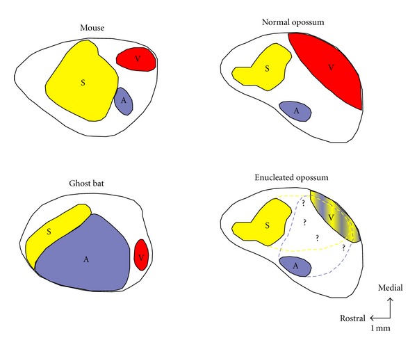
The relative positioning of the primary sensory areas is remarkably consistent across mammalian species. Visual cortex, for example, is always posterior: There is no mammal with visual cortex at the front of the brain. This points to a deep and reliably reconstructed pattern in how cortical areas are arranged, one conserved across the whole radiation of mammals. If it is a “plan”, it is one that the developmental process rebuilds in each generation.
The figure below shows how in mice, ghost bats, and opossums, primary sensory areas occupy broadly similar relative positions, but the area devoted to touch, hearing, or vision differs with the animal’s way of life. Note the size of auditory cortex in the ghost bat, which echolocates. The fourth panel shows an opossum deprived of vision early in development. In the original study, the eyes were removed on postnatal day 4, before thalamocortical afferents entered the cortex. In adulthood, cortical territory normally devoted to vision responded to auditory and somatosensory stimulation (Kahn & Krubitzer, 2002). Sensory input during development thus also shapes the details of cortical organization.
To understand these differences, start with the idea that within each primary sensory area the cortex contains an orderly spatial representation of the receptor surface it serves. Primary visual cortex contains a map of the retina; primary auditory cortex contains a map organized by sound frequency; primary somatosensory cortex contains a somatotopic map of the body surface, in which neighboring body parts are represented by neighboring cortical regions. These maps preserve aspects of the spatial relationships across receptor surfaces, although cortical magnification and discontinuities mean that they are not literal copies of the body’s physical layout.
As we saw in Evolution and Development, the bat wing and the mouse paw are two developmental trajectories from a shared ancestral limb. The Prx1 experiment discussed there shows one way that altered gene regulation can affect limb growth. Differences in limb form and use can, in turn, enter into the activity through which cortical maps develop. To understand the cortical map, we need to follow how the brain develops in relation to the body and its use.
These cross-species comparisons tell us that cortical maps reflect the bodies animals have and the ways of life those bodies make possible. But the idea that cortical maps “reflect” or “represent” bodies can be misleading, because it suggests a one-way process in which the body is a certain way and the cortex mirrors it.
The developing cortex is being organized by patterns of activity and connectivity that arrive through developing pathways, and it is also already being patterned from within, by molecular gradients that shape its regional structure before any of that activity arrives. The body is developing at the same time as the cortex, not before it, and the activity that links them runs through structures, such as the thalamus and the sensory pathways, that are themselves still being built.
Neither the developing body nor the developing brain instructs the other. They emerge together, each shaping the conditions for the other’s further development. So the question of how cortical areas become specialized is a question about how multiple developing systems, the body, the cortex, and the pathways between them, come to be organized in relation to each other and to the way of life of the organism.
Two Models of Cortical Development
The question of how cortical areas develop has often been framed around two contrasting models. One emphasizes intrinsic molecular factors, the other the role of neural activity delivered to the cortex from elsewhere. Both models are worth understanding, and so are the limits of setting them against each other, because the contrast repeats an old problem in the study of development: treating development as a contest between two separate sources.
First, some background. Sensory information from the body and the sense organs does not arrive at the cortex directly. Signals are relayed through the thalamus, a collection of nuclei deep in the brain. Different thalamic nuclei handle different modalities: the lateral geniculate nucleus (LGN) relays visual information from the retina to primary visual cortex, the medial geniculate nucleus (MGN) relays auditory information to primary auditory cortex, and the ventral posterior nucleus relays somatosensory information to primary somatosensory cortex. These thalamocortical connections are a major pathway by which patterns of neural activity reach the developing cortex.
The protomap model, associated with the work of Pasko Rakic and colleagues (2009), states that the fate of cortical areas is determined very early in development by intrinsic molecular factors, before thalamocortical connections have arrived. The protocortex model, associated with the work of Dennis O’Leary (1989), emphasizes the initially less differentiated character of cortical tissue and the contribution of afferent input to the emergence of distinct cortical areas. In this view, a cortical region’s specialization depends partly on the input it receives, not just on where it is located.
Support for the protomap model comes from research on patterning centers in the developing telencephalon (Sansom & Livesey, 2009). These centers secrete signaling molecules (morphogens) that diffuse across the ventricular zone, creating concentration gradients. Through their influence on transcription factors, such as those coded by the Pax6 and Emx2 genes, these gradients drive a graded pattern of gene expression within the early cortical primordium, influencing neural progenitor cells to give rise to region-specific neurons with distinct molecular, physiological, and connectivity features. Note that we are talking about gradients of expression, not a simple on/off pattern. The identity of a cortical region emerges from the relative concentrations of multiple transcription factors at a given location.
Jernigan and Stiles (2017) provide a useful summary of how these factors interact. During neurogenesis, molecular signaling within the ventricular zone influences the initial “areal fate” of cortical projection neurons, in terms of whether they will migrate toward motor, visual, or auditory areas. But, as they put it, “this very early fate specification is primitive and malleable” (Jernigan & Stiles, 2017, p. 5). The full differentiation of a neuron as a motor or visual neuron depends on many subsequent developmental events, involving both continued molecular signaling and patterns of neural activity.
Although there is strong evidence for the molecular gradients the protomap describes, a question remains. If the origins of cortical regions can be found in the activity of patterning centers and intrinsic gradients of gene expression, then the origin of those gradients cannot itself be the activity of other patterning centers or other gradients, because that would simply push the question back a step. Nor is it enough to say that the intrinsic activity of patterning centers is “genetically specified” and therefore needs no further explanation. We are still left with some explaining to do, and we will come back to it at the end of the reading.
As Kast and Levitt (2019) put it, “there is conclusive support for the protomap hypothesis, yet it is unlikely to be so simple” (p. 79). They review extensive evidence that thalamic input influences cortical development at several levels. In practice, the field has moved toward seeing cortical development as involving both intrinsic molecular patterning and activity-dependent mechanisms. Larger-scale regions may be laid down by molecular gradients, while finer-scale organization within those regions arises through neural activity. Part of the difficulty in separating the two is experimental: in human development, projections from the thalamus are present in the cortical neuroepithelium even before neurogenesis begins, which makes it hard to tease apart their contribution from that of intrinsic patterning (Cadwell et al., 2019, p. 992).
Pitting protomap against protocortex, in the end, is a little like pitting nature against nurture. We could say it is some of both, or some kind of interaction, but this has the same limitation, because it assumes a separation that may not actually exist. A more interesting question is what kinds of activity are involved in the development of the cortex, and where that activity comes from.
Whisker Barrel Fields
One of the best-studied examples of how cortical maps are shaped comes from research on the barrel fields in rodent somatosensory cortex. Rodents rely heavily on their whiskers (vibrissae) for sensing the world, using them to navigate in the dark, to identify objects by texture and shape, and to detect air currents. Classic research in the 1970s showed how each whisker is represented by a discrete cluster of neurons in primary somatosensory cortex, and together these clusters form a map of the whisker array on the face. The clusters are called “barrels” because of their distinctive shape in cross-section.
The pathways supporting the barrel map begin developing prenatally, while the recognizable cortical barrels emerge during the first days after birth. Van der Loos and Woolsey (1973) showed that injury to a whisker follicle at birth can prevent the corresponding cortical barrel from developing normally. Comparable injury after the first few postnatal days no longer produces the same rearrangement of the barrel pattern. This is a sensitive period for that anatomical organization, rather than a point after which the cortex ceases to be plastic. Damage to the follicle also differs from simply trimming a whisker and reducing its use (Erzurumlu & Gaspar, 2012).
The barrels show that patterned input from the whiskers, relayed via the thalamus, organizes the detailed structure of the cortical map. But the barrels form within a region that has already been established as somatosensory cortex rather than visual or auditory cortex. Something determined the broad identity of this cortical region before the whisker input arrived to refine it. The activity from the whiskers shapes the map, but it does not create the somatosensory region from scratch. This is the picture the field has converged on: the protomap establishes that a particular region of cortex will be somatosensory, and the detailed organization of the body map within that region depends on patterns of activity coming from the body.
Activity in the Developing Brain
When we say that neural activity shapes cortical development, we might picture sensory input from the outside world arriving at the cortex and molding it into shape. That picture is not wrong, but much of the activity that shapes early cortical development is generated within the developing nervous system itself.
So we need to think about neural activity in two ways. One kind is intrinsic, spontaneous activity that does not seem to be connected to sensory input from outside the organism. The other involves activity in the pathways from the sense organs, such as from the whiskers. Both turn out to be important, and both complicate the distinction between what is “intrinsic” and what is “extrinsic” to cortical development.
Retinal waves
Before the retina supports mature patterned vision, waves of spontaneous electrical activity sweep across its surface. Retinal waves have been studied most extensively in rodents, in which they occur in the days after birth, before the eyes open, including in vivo work showing their propagation through the developing visual system (Ackman et al., 2012). In macaques, retinal waves have been recorded during fetal development, well before birth (Warland et al., 2006).
The waves are not random. Neighboring retinal cells tend to fire together, and the waves propagate across the retina in a coordinated, spatially organized pattern. Because neighboring retinal cells tend to be active together, the activity transmitted along the developing optic pathway carries spatial information about the structure of the retina, that is, about which cells are near each other. This activity is relayed through the thalamus (specifically the LGN) to the developing visual cortex, where it helps establish the topographic mapping between retina and cortex, so that neighboring points on the retina come to be represented by neighboring neurons in the cortex.
Experimental work also implicates retinal activity in the segregation of inputs from the two eyes, although the details differ across species and between the thalamus and cortex. Disrupting retinal waves can alter the refinement of visual connections (Ackman & Crair, 2014).
So the visual system is being organized by activity that the developing retina generates on its own, without any visual input from the world, in ways that contribute to the organization through which the animal will later see. As vision develops, visually driven activity becomes increasingly important, and the system continues to be refined within an organization that earlier activity helped establish. This transition occurs after birth in rodents and on a different timetable in more mature newborns, so birth itself is not a universal dividing line.
There is a connection here to Gilbert Gottlieb’s (1980) research on ducklings. The duckling’s preference for its species’ maternal call turned out to depend on prenatal exposure to the duckling’s own vocalizations. What looked like a hardwired species-typical behavior was actually maintained by self-generated experience that nobody had thought to look for until someone disrupted it. Retinal waves are a similar kind of finding at the neural level. The developing nervous system supplies some of the activity through which it becomes organized. Enacting Life takes up the broader implications of this active role.
Body maps and fetal movement
Primary somatosensory cortex contains an orderly representation of the body surface, with neighboring body parts represented by neighboring cortical regions. Somatotopic organization is detectable even in preterm infants, showing that it begins to take shape before the usual time of birth (Dall’Orso et al., 2018).
The fetus is not a passive occupant of the womb. Especially in the later part of pregnancy, a fetus moves, kicks, stretches, brings its hand to its face, and touches the uterine wall. These movements generate sensory consequences. Muscles, joints, and skin are activated, producing patterned neural activity that reaches the developing somatosensory cortex via the thalamus. Because body parts that are physically close tend to move together, so that when the arm moves the hand moves with it, the activity arriving at the cortex carries spatial information about the body’s structure.
The proposal is that the same logic as retinal waves operates in a different system: correlated activity from neighboring parts of a receptor surface helps establish an orderly cortical map. Much of the direct evidence comes from newborn rats, in which spontaneous limb twitches drive bursts of activity in the corresponding parts of somatosensory cortex (Khazipov et al., 2004).
Back to the Developmental System
The same principle that operates in whisker barrels, retinal waves, and body maps, namely that correlated activity from a spatially structured receptor surface establishes an orderly cortical map, offers a way to investigate the development of other sensory maps. Different animals have different bodies, those bodies have different receptor surfaces, and those surfaces generate different patterns of spatially structured activity during development.
Consider the bat wing again. The wing membrane is a large, continuous sensory surface. The proposal is that activity generated across this surface helps organize its cortical representation, alongside molecular patterning and the development of sensory pathways. The wing’s size, innervation density, and spatial structure would therefore be part of the conditions under which its map in S1 develops. It is a hypothesis that follows from the whisker and retinal-wave work, not something those studies tested.
What comes first, the body part or the cortical area that represents it? The answer is neither, because the body and the cortex develop at the same time. The protomap is laying down broad cortical regions through molecular gradients before thalamocortical connections have arrived to carry information from the body. Meanwhile, limb buds are growing, innervation patterns are being established, and the thalamic nuclei that will relay signals between body and cortex are forming their own connections. These processes overlap in time. The cortex is not waiting for the body to be finished before mapping it, and the body is not waiting for the cortex to be ready before connecting to it. Body and brain develop together, constraining each other as they go.
The same holds for their evolutionary history. The bat wing is not an invention that the cortex then had to accommodate. The wing and its cortical representation emerged together, as part of the same evolving life cycle. Evolutionary changes in the wing occurred within a developmental system that also produced its innervation and cortical representation.
The Prx1 experiment fits this picture. Swapping in the bat version of the enhancer lengthened mouse forelimbs modestly, and only for a while, within a developmental process that was already organized (Cretekos et al., 2008). It did not specify a wing, and the study did not look at cortical maps.
This is also why brain development is reliable without being genetically programmed. The reliability comes not from instructions in DNA but from the reliable reconstruction of the whole developmental system, generation after generation. The same molecular gradients, the same kinds of spontaneous activity, the same thalamocortical pathways, and the same relationship between body and cortex are reproduced each time a new organism develops, because the conditions for their development are themselves reliably present. DNA is part of what gets reliably reconstructed, but so is the cell, the uterine environment, the body plan, and the physics of how layered sheets of tissue fold and grow. These recurring conditions help explain why cortical organization is reliable. Differences in them help us investigate why an individual develops a particular map.
What Organizes the Organizer?
Earlier we met the “organizers” that Spemann and Mangold identified, regions of embryonic tissue that induce the cells around them to develop in particular ways. It is natural to ask where an organizer comes from, and following that question is instructive.
Much of what we know comes from work in the frog Xenopus, a common model organism in developmental biology. When researchers traced the origin of the organizer, they found that it was induced by a neighboring region of the embryo. That region, in turn, was established by the redistribution of signaling molecules already present in the egg, triggered by the entry of the sperm at fertilization. The relevant maternal molecules had accumulated or become localized during the egg’s formation in the mother’s body (Houston, 2017).
Many of these steps have been characterized experimentally, and at no point in the chain do you find a set of instructions that specifies the outcome. At each step, an existing structure changes the conditions for the next step.
The same lesson appears if we look at the neural tube. Can we say there is a gene responsible for making one side dorsal and the other ventral? All cells have the same DNA, so any such gene is present in every part of the tube. We can say there are gradients of gene expression that influence dorsal and ventral cell identities. But is there a gene that determines the gradients? The cycle of questions just keeps going back.
If you keep asking what came before, you end up at how the egg was built in the mother’s body, which depends on her development, which depended on the same kind of cascade in the previous generation. There is no first step, and no unmoved mover at the end of the questions. There is a cycle that is reliably reconstructed each time, and the reliability comes not from a program being executed but from the conditions for each step being dependably present.
There are always living cells flowing through the cycle of lives, so some cellular structure is always preexisting, though not in a genetic sense. What we need in place of a blueprint is the idea of stable developmental processes that involve genes but are not directed by them.
Sources
Ackman, J. B., Burbridge, T. J., & Crair, M. C. (2012). Retinal waves coordinate patterned activity throughout the developing visual system. Nature, 490, 219–225. https://doi.org/10.1038/nature11529
Ackman, J. B., & Crair, M. C. (2014). Role of emergent neural activity in visual map development. Current Opinion in Neurobiology, 24, 166–175. https://doi.org/10.1016/j.conb.2013.11.011
Cadwell, C. R., Bhaduri, A., Mostajo-Radji, M. A., Keefe, M. G., & Nowakowski, T. J. (2019). Development and arealization of the cerebral cortex. Neuron, 103(6), 980–1004. https://doi.org/10.1016/j.neuron.2019.07.009
Cretekos, C. J., Wang, Y., Green, E. D., Martin, J. F., Rasweiler, J. J., IV, & Behringer, R. R. (2008). Regulatory divergence modifies limb length between mammals. Genes & Development, 22(2), 141–151. https://doi.org/10.1101/gad.1620408
Dall’Orso, S., Steinweg, J. K., Allievi, A. G., Edwards, A. D., Burdet, E., & Arichi, T. (2018). Somatotopic mapping of the developing sensorimotor cortex in the preterm human brain. Cerebral Cortex, 28(7), 2507–2515. https://doi.org/10.1093/cercor/bhy050
Darnell, D., & Gilbert, S. F. (2017). Neuroembryology. WIREs Developmental Biology, 6(1), e215. https://doi.org/10.1002/wdev.215
Desgent, S., & Ptito, M. (2012). Cortical GABAergic interneurons in cross-modal plasticity following early blindness. Neural Plasticity, 2012, 590725. https://doi.org/10.1155/2012/590725
Erzurumlu, R. S., & Gaspar, P. (2012). Development and critical period plasticity of the barrel cortex. European Journal of Neuroscience, 35(10), 1540–1553. https://doi.org/10.1111/j.1460-9568.2012.08075.x
Gottlieb, G. (1980). Development of species identification in ducklings: VI. Specific embryonic experience required to maintain species-typical perception in Peking ducklings. Journal of Comparative and Physiological Psychology, 94(4), 579–587. https://doi.org/10.1037/h0077691
Houston, D. W. (2017). Vertebrate axial patterning: From egg to asymmetry. In F. Pelegri, M. Danilchik, & A. Sutherland (Eds.), Vertebrate development: Maternal to zygotic control (pp. 209–306). Springer. https://doi.org/10.1007/978-3-319-46095-6_6
Jernigan, T. L., & Stiles, J. (2017). Construction of the human forebrain. WIREs Cognitive Science, 8(1–2), e1409. https://doi.org/10.1002/wcs.1409
Kahn, D. M., & Krubitzer, L. (2002). Massive cross-modal cortical plasticity and the emergence of a new cortical area in developmentally blind mammals. Proceedings of the National Academy of Sciences, 99(17), 11429–11434. https://doi.org/10.1073/pnas.162342799
Kast, R. J., & Levitt, P. (2019). Precision in the development of neocortical architecture: From progenitors to cortical networks. Progress in Neurobiology, 175, 77–95. https://doi.org/10.1016/j.pneurobio.2019.01.003
Khazipov, R., Sirota, A., Leinekugel, X., Holmes, G. L., Ben-Ari, Y., & Buzsáki, G. (2004). Early motor activity drives spindle bursts in the developing somatosensory cortex. Nature, 432, 758–761. https://doi.org/10.1038/nature03132
Kostović, I., & Rakic, P. (1990). Developmental history of the transient subplate zone in the visual and somatosensory cortex of the macaque monkey and human brain. Journal of Comparative Neurology, 297(3), 441–470. https://doi.org/10.1002/cne.902970309
Lumen Learning. (n.d.). Embryonic development. In Anatomy and Physiology II. https://courses.lumenlearning.com/suny-ap2/chapter/embryonic-development/
O’Leary, D. D. M. (1989). Do cortical areas emerge from a protocortex? Trends in Neurosciences, 12(10), 400–406. https://doi.org/10.1016/0166-2236(89)90080-5
Rakic, P., Ayoub, A. E., Breunig, J. J., & Dominguez, M. H. (2009). Decision by division: Making cortical maps. Trends in Neurosciences, 32(5), 291–301. https://doi.org/10.1016/j.tins.2009.01.007
Sansom, S. N., & Livesey, F. J. (2009). Gradients in the brain: The control of the development of form and function in the cerebral cortex. Cold Spring Harbor Perspectives in Biology, 1(2), a002519. https://doi.org/10.1101/cshperspect.a002519
Spemann, H. (1935). The organizer-effect in embryonic development. Nobel Lecture, December 12, 1935. NobelPrize.org. https://www.nobelprize.org/prizes/medicine/1935/spemann/lecture/
Spemann, H., & Mangold, H. (2001). Induction of embryonic primordia by implantation of organizers from a different species (V. Hamburger, Trans.). The International Journal of Developmental Biology, 45(1), 13–38. https://ijdb.ehu.eus/article/11291841 (Original work published 1924).
Van der Loos, H., & Woolsey, T. A. (1973). Somatosensory cortex: Structural alterations following early injury to sense organs. Science, 179(4071), 395–398. https://doi.org/10.1126/science.179.4071.395
Warland, D. K., Huberman, A. D., & Chalupa, L. M. (2006). Dynamics of spontaneous activity in the fetal macaque retina during development of retinogeniculate pathways. The Journal of Neuroscience, 26(19), 5190–5197. https://doi.org/10.1523/JNEUROSCI.0328-06.2006
Explore Further
For an introductory account of early embryonic development, see Lumen Learning (n.d.) in Sources.