3.3 Plasticity and Constraint
The cortex gives us a way to examine a question that runs through this resource: how can development produce outcomes that are both reliable and variable? Across mammals, the primary sensory areas occupy similar relative positions. Their detailed organization, however, differs across species and individuals as the cortex develops in relation to a particular body and its activity. Plasticity, the capacity for change, is part of this developmental process. The question is how far that capacity extends and what constrains it.
Because the cortex is still being organized after birth, it is a system we can probe: altering the activity that reaches it is one of the best ways to see how it is put together and where its limits lie. This reading considers what happens when sensory input is missing or redirected, when the body has an unusual structure, and when input to an established map is lost. Comparing the cases lets us examine how the response depends on when the change occurs and what is already established.
The Space of Possibility Narrows
In many developmental processes, the range of possible outcomes narrows as organization becomes established. Spemann’s transplantation experiments with newt embryos provide a clear example (Spemann, 1935). Early in gastrulation, transplanted cells developed according to their new location. Later, they continued along the course established before transplantation. Earlier interactions had changed how the cells responded to their surroundings, constraining what subsequent interactions could do.
Those were, in effect, perturbation experiments, and what they showed is that the response to a perturbation depends on the state of the system when it occurs. That point runs through everything that follows.
Development can also open new possibilities as new capacities emerge, so narrowing is not a universal rule. In sensory development, however, there are well-studied windows during which particular kinds of activity have especially strong effects. Vision and hearing provide two examples.
Most of the evidence considered here concerns sensory and motor regions of the cortex. These offer useful ways to study plasticity, but their development should not be treated as a model for every cortical region or every psychological capacity.
Vision and Hearing
Dense cataracts present at birth can block patterned light from reaching the retina. If they are not removed early, visual difficulties can persist after surgery (Lewis & Maurer, 2005). Cataracts that develop in adulthood can often be treated successfully because the visual system has already developed with patterned input. The comparison shows why restoring input is not always enough: its developmental effects depend on when it becomes available.
Research by David Hubel and Torsten Wiesel helped explain this timing effect (Wiesel & Hubel, 1963). When they sutured one eyelid shut in young kittens, depriving that eye of patterned visual input, far fewer neurons in visual cortex responded to the deprived eye. Responses were dominated by the other eye, and the effects could persist after the closed eye was reopened. Comparable deprivation in adult cats had much smaller effects. These findings helped establish the idea of a critical period, a window of heightened sensitivity to particular conditions. Hubel and Wiesel’s work on visual processing, including ocular dominance, later earned them the Nobel Prize.
As discussed in Brain, Body, World, visual organization begins before patterned vision is available. Retinal waves, spontaneous activity generated by the developing retina, help establish the orderly mapping of retinal locations onto the brain. Later visual input refines this developing system, including the coordination of signals from the two eyes. Different visual properties have different developmental histories and do not all depend on experience in the same way. When a critical period closes, certain changes become harder to produce, but the visual system goes on developing and changing.
Timing is also important in hearing. Cochlear implants convert signals from a microphone into electrical stimulation of the auditory nerve. Among children born deaf, earlier implantation is generally associated with better auditory and spoken-language outcomes, although outcomes vary and later implantation can still be beneficial (Kral & Sharma, 2012). Here too, the effect of restored input depends partly on the developmental history of the system receiving it.
For both vision and hearing, the space of possibilities is wide early in development and then narrows. Missing input during these periods can have lasting consequences, even though other forms of change remain possible later.
Rewiring the Developing Brain
Could a developing cortical area respond to a different sensory modality if its input were redirected? This question bears on the protomap and protocortex models discussed in the previous reading. Mriganka Sur and colleagues at MIT investigated it through experiments with newborn ferrets.
The researchers redirected retinal input toward what would normally become auditory cortex. Sharma, Angelucci, and Sur (2000) examined the resulting cortical organization. In related behavioral experiments, von Melchner and colleagues (2000) found that ferrets could use the altered pathway to distinguish visual from auditory stimuli.
Ferrets were suitable for these experiments because their visual connections are relatively immature at birth. Normally, auditory signals travel from the cochlea through the inferior colliculus (IC) to the medial geniculate nucleus (MGN) of the thalamus, which projects to primary auditory cortex. Neonatal surgery disrupted this auditory input and removed the superior colliculus, one of the retina’s normal targets. Retinal axons could then innervate the MGN. Because the MGN still projected to auditory cortex, retinal activity reached cortex through an altered pathway.
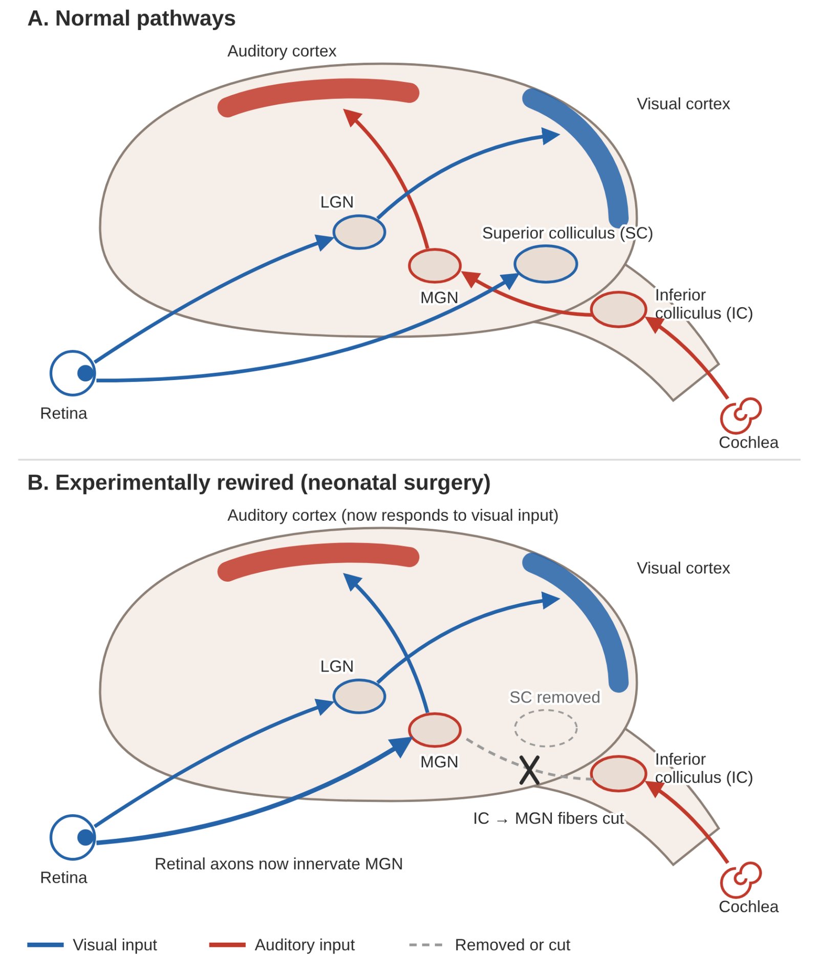
Neurons in the rewired auditory cortex showed orientation selectivity: they responded preferentially to edges at particular angles. These preferences were arranged in pinwheel-like patterns resembling those found in visual cortex.
The altered input had shaped cortical organization, but the rewired area did not become identical to typical visual cortex. One way to read this is that a cortical region is best suited to certain kinds of input but can still work with input from another modality, though not in the same way. The space of possibilities is structured: not every region is equally suited to every input. We return below to how these experiments should be interpreted.
The Possibility Space and the Body
Body maps in primary somatosensory cortex offer another way to investigate plasticity. Their organization begins before birth. As discussed in Brain, Body, World, one proposal is that activity generated by fetal movement contributes to this organization, as correlated sensory activity helps developing pathways become spatially ordered.
What happens when the body differs from the typical one? If cortical maps develop through activity involving the body, we should expect them to reflect the body that is present.
Polydactyly is the development of additional fingers or toes. Digit number takes shape during embryonic limb development through interactions involving signaling molecules such as Sonic hedgehog (Shh). Changes in these interactions can produce extra digits, whose structure and function vary across cases.
Mehring and colleagues (2019) studied two people with preaxial polydactyly, each with six fully developed fingers on each hand. The additional finger lay between the thumb and index finger and had its own muscles, tendons, and nerves.
High-resolution fMRI revealed a distinct sensorimotor representation of the additional finger. It could move independently, and participants coordinated it with their other fingers during manipulation. They could perform some tasks with one hand that five-fingered participants needed two hands to accomplish.
The additional finger was integrated into both cortical organization and skilled action. The same developmental process that maps a five-fingered hand, a bat wing or a mouse paw had mapped a six-fingered one. In Freaks of Nature, Mark Blumberg (2009) discusses other bodily differences that reveal the flexibility of development, including Johnny Eck, a performer born without the lower half of his body. His case illustrates how a functional way of life can develop through relations among an unusual body, its activity, and its surroundings.
Tucciarelli and colleagues (2026) examined a complementary case: children and adults born without one hand. The cortical territory that would typically represent the missing hand was more responsive to other body parts. This pattern was already present in children aged five to seven and was similar in adults, despite differences in their compensatory behaviors. The comparison suggests that substantial features of this map are established early, although it does not track the same individuals from childhood into adulthood.
From Individual Development to Species Differences
The relation between body and cortical map also helps us understand differences across species. A bat’s wing, a platypus’s bill, and a mouse’s whiskers each participate in the development of distinctive cortical representations. Shared principles of activity-dependent development can produce different outcomes because the bodies and their activity differ. The bodies themselves also develop through processes reconstructed across generations, with genes among the resources involved. In some evolutionary cases, developmental plasticity can precede and channel subsequent genetic change (West-Eberhard, 2003).
Polydactyly shows that a substantial bodily difference can arise within a single generation: an additional digit can develop with muscles, nerves, and a cortical representation. Producing such a variant is a different question from explaining its evolutionary persistence. Six-fingered hands have not become typical in humans as elongated digits have in bats. What takes evolutionary time is not the appearance of a variant but the stabilization of a way of life around it. Why that has not happened for six fingers would depend on functional advantages, costs elsewhere in development, and population history.
A Side Note on Polydactyly
Different forms of polydactyly have different developmental consequences. One route to preaxial polydactyly involves a regulatory change in the limb-specific enhancer controlling Sonic hedgehog expression. This can cause Shh activity in an unusual location within the limb bud without changing the Shh protein itself (Maas & Fallon, 2005). Polydactyly can also occur within a broader syndrome. Ellis-van Creveld syndrome is associated with variants in EVC or EVC2, whose products participate in ciliary Hedgehog signaling (Blair et al., 2011). Extra digits can occur alongside short limbs, heart defects, and dental anomalies because the affected processes operate across several tissues.
Ellis-van Creveld syndrome is unusually common among the Old Order Amish of Lancaster County. This reflects a founder variant and a history of relative reproductive isolation and marriage within the community (McKusick, 2000), rather than evidence that natural selection favored the variant. The community’s way of life stabilized a population structure, and the variant came along for the ride. The bat wing, by contrast, illustrates the incorporation of developmental variation into an evolving way of life.
An additional finger can become part of skilled action, and useful patterns of activity can develop around a missing body part. Other developmental alterations affect several tissues and carry substantial costs. The capacity to accommodate variation depends on which processes are altered and how those alterations affect the organism as a whole.
When Input to Maps Is Disrupted
The cases above involve bodies and maps developing together. What happens when input to an established adult map is lost? In the early 1980s, Michael Merzenich and colleagues mapped the hand representation in adult monkeys’ primary somatosensory cortex. As in Penfield’s work with humans, they related responses at different cortical locations to stimulation of different body parts.
After amputating a middle finger, Merzenich and colleagues (1984) remapped the cortex several months later. Locations that had responded most strongly to that finger now responded to neighboring fingers. This was widely interpreted as adjacent representations taking over the deprived territory. This was not supposed to happen if you took Hubel and Wiesel’s findings as the general rule: the map had changed in adulthood, well past anything that could be called a critical period. What kind of change had occurred, however, remained a question.
Pons and colleagues (1991) studied adult monkeys whose sensory input from one arm had been surgically eliminated years earlier. Stimulation of the face evoked responses in somatosensory territory previously associated with the arm. These findings helped inspire V. S. Ramachandran’s work on phantom limbs in humans.
These animal studies involved severe alterations to the body or its sensory pathways, alongside direct recording from neural tissue. Their invasiveness is part of the history of this research and needs to be considered when evaluating it. Ethical oversight has changed substantially since many of the foundational studies were conducted.
Related findings come from blindness and deafness in humans. During Braille reading, blind participants show activity in cortical areas usually associated with vision, with more extensive activity in those who became blind early (Burton et al., 2002). In deaf participants, regions usually associated with auditory processing can respond to visual stimuli (Bola et al., 2017). Such responses are described as cross-modal because they involve a different sensory modality from the one typically associated with that region.
These observations are often taken to mean that cortical regions have acquired new functions. Before considering that interpretation, it helps to distinguish changes associated with early developmental conditions from those that occur through an individual’s later experience.
Experience-Expectant and Experience-Dependent Plasticity
Greenough and colleagues (1987) distinguished experience-expectant from experience-dependent plasticity. Experience-expectant processes depend on conditions ordinarily encountered during a species’ development, such as patterned visual input during an early window. Experience-dependent changes reflect an individual’s particular history and can occur throughout life. Training-related differences in skilled string musicians, changes associated with enriched environments in rodents, and the adult map changes discussed above illustrate this second category. The term “expectant” is shorthand for a developmental dependency; it does not mean that the system literally anticipates the input.
The distinction describes when experience has its effects, while leaving us to explain how those effects occur. It is better read as two ways in which a single activity-dependent process can be structured in time than as two separate mechanisms. Both concern how neural organization changes through activity, including spontaneously generated activity and responses to the surroundings. Their timing and consequences depend on the processes involved.
It is tempting to use accounts of extreme deprivation, such as the child known as “Genie” or so-called feral children, as human parallels to the kitten studies. But these cases cannot establish critical periods. Their circumstances are confounded, and deprivation is difficult to separate from other conditions. Language also develops through processes that cannot simply be equated with the development of sensory maps. Questions about sensitive periods for language require systematic research, rather than conclusions drawn from isolated cases.
Reframing Plasticity
In a 2023 review paper, Tamar Makin and John Krakauer question whether changes in cortical maps establish that regions have acquired new functions. They emphasize the strengthening or unmasking of pre-existing connections. In the finger-amputation studies, for example, responses to neighboring fingers could already be present within the territory most responsive to the missing finger. Losing the dominant input could make these weaker responses more prominent. Subsequent activity could further strengthen or refine them. This is plasticity, but it need not involve a cortical region acquiring an entirely new capacity. They apply related reasoning to the findings of Pons and colleagues.
Makin and Krakauer also reconsider the ferret experiments. The altered pathway supported visual behavior, but the behavioral task demonstrated detection of visual stimuli rather than use of the new orientation map. They argue that shared properties of visual and auditory cortex could support processing of the redirected input. The experiments reveal substantial flexibility under unusual conditions, but do not establish unrestricted capacity to acquire new functions.
For blindness and deafness, the same question applies: does a stronger response to another modality establish a new function? Some cortical regions already respond to several modalities. Analyses that compare patterns of activity across stimuli, including representational similarity analysis, can reveal similarities in how a region organizes those responses across groups. A change in the source or strength of input therefore need not imply a change in the kind of processing performed. Whether that holds has to be judged region by region and task by task.
A related possibility has been proposed for synesthesia, in which one experience consistently accompanies another, such as a color experienced when hearing a sound. Grossenbacher and Lovelace (2001) proposed that reduced inhibition of feedback between cortical regions could contribute to synesthetic experience. This resembles the unmasking interpretation, though it remains a hypothesis.
How researchers construct a cortical map also affects what an apparent change in that map means. Many maps use a “winner-takes-all” procedure. Each cortical location is labeled according to the tested stimulus that produces the strongest response there, such as a particular finger or eye. The resulting boundaries can look clear-cut even when responses overlap. A location labeled “index finger” may also respond to stimulation of other fingers, just less strongly.
Ocular dominance provides an example. Neurons can respond to both eyes while responding more strongly to one. The crisp alternating columns you might learn about in a neuroscience class summarize those preferences. The columns are real, but their sharp borders come from how the map is drawn, and not every neuron in a column responds only to one eye.
If the strongest input is removed, another input may become the winner. A newly drawn map will then assign that location a different label. This can look like one representation has taken over another, even if the neurons already responded to both inputs. The map has changed, but the change alone does not tell us whether new connections formed, existing connections strengthened, or previously weak responses became easier to detect.
For Makin and Krakauer, a change in the measured map does not by itself establish a new mechanism or function. Demonstrating a new functional capacity requires more than showing that a cortical location responds most strongly to a different stimulus.
Plasticity, Constraints, and the Developmental System
Altered input can bring previously weak responses into prominence without the cortex acquiring a new function. Existing structure and connections constrain what can change. But that leaves a developmental question: how did those constraints arise?
In their abstract, Makin and Krakauer describe the constraints in terms of a structural “blueprint,” in quotation marks. In their introduction, they characterize the reorganization view as assuming that experience can override “the genetic blueprint of brain function.” Even as framing, this language suggests that genes specify an organization which experience can only modify. There is no genetic blueprint. The developmental account pursued in this resource asks instead how that organization is produced. Genes participate within the developing system; their role depends on what else is happening there. Calling the result a blueprint risks treating the organization that development achieves as an explanation for how it was achieved.
A developmental explanation can recognize strong constraints without locating them in a genetic plan. Processes stabilized over evolutionary time reliably produce particular structures and connections. As these develop, they shape the effects of subsequent activity. The resulting limits are real, but they are limits of a developing system with a history.
Plasticity and constraint are therefore linked throughout development. The same processes that allow a body map to accommodate an additional finger also establish connections that make some later changes possible and others difficult. After input is lost, existing pathways may support a different pattern of responses. Explaining that flexibility requires us to ask both what the system can do now and how it developed the organization that makes those responses possible. Plasticity, understood this way, is movement within a structured space rather than unlimited malleability.
Sources
Blair, H. J., Tompson, S., Liu, Y.-N., Campbell, J., MacArthur, K., Ponting, C. P., Ruiz-Perez, V. L., & Goodship, J. A. (2011). Evc2 is a positive modulator of Hedgehog signalling that interacts with Evc at the cilia membrane and is also found in the nucleus. BMC Biology, 9, 14. https://doi.org/10.1186/1741-7007-9-14
Blumberg, M. S. (2009). Freaks of nature: What anomalies tell us about development and evolution. Oxford University Press.
Bola, Ł., Zimmermann, M., Mostowski, P., Jednoróg, K., Marchewka, A., Rutkowski, P., & Szwed, M. (2017). Task-specific reorganization of the auditory cortex in deaf humans. Proceedings of the National Academy of Sciences, 114(4), E600–E609. https://doi.org/10.1073/pnas.1609000114
Burton, H., Snyder, A. Z., Conturo, T. E., Akbudak, E., Ollinger, J. M., & Raichle, M. E. (2002). Adaptive changes in early and late blind: A fMRI study of Braille reading. Journal of Neurophysiology, 87(1), 589–607. https://doi.org/10.1152/jn.00285.2001
Greenough, W. T., Black, J. E., & Wallace, C. S. (1987). Experience and brain development. Child Development, 58(3), 539–559. https://doi.org/10.2307/1130197
Grossenbacher, P. G., & Lovelace, C. T. (2001). Mechanisms of synesthesia: Cognitive and physiological constraints. Trends in Cognitive Sciences, 5(1), 36–41. https://doi.org/10.1016/S1364-6613(00)01571-0
Kral, A., & Sharma, A. (2012). Developmental neuroplasticity after cochlear implantation. Trends in Neurosciences, 35(2), 111–122. https://doi.org/10.1016/j.tins.2011.09.004
Lewis, T. L., & Maurer, D. (2005). Multiple sensitive periods in human visual development: Evidence from visually deprived children. Developmental Psychobiology, 46(3), 163–183. https://doi.org/10.1002/dev.20055
Maas, S. A., & Fallon, J. F. (2005). Single base pair change in the long-range Sonic hedgehog limb-specific enhancer is a genetic basis for preaxial polydactyly. Developmental Dynamics, 232(2), 345–348. https://doi.org/10.1002/dvdy.20254
Makin, T. R., & Krakauer, J. W. (2023). Against cortical reorganisation. eLife, 12, e84716. https://doi.org/10.7554/eLife.84716
McKusick, V. A. (2000). Ellis-van Creveld syndrome and the Amish. Nature Genetics, 24, 203–204. https://doi.org/10.1038/73389
Mehring, C., Akselrod, M., Bashford, L., Mace, M., Choi, H., Blüher, M., Buschhoff, A.-S., Pistohl, T., Salomon, R., Cheah, A., Blanke, O., Serino, A., & Burdet, E. (2019). Augmented manipulation ability in humans with six-fingered hands. Nature Communications, 10, 2401. https://doi.org/10.1038/s41467-019-10306-w
Merzenich, M. M., Nelson, R. J., Stryker, M. P., Cynader, M. S., Schoppmann, A., & Zook, J. M. (1984). Somatosensory cortical map changes following digit amputation in adult monkeys. Journal of Comparative Neurology, 224(4), 591–605. https://doi.org/10.1002/cne.902240408
Pons, T. P., Garraghty, P. E., Ommaya, A. K., Kaas, J. H., Taub, E., & Mishkin, M. (1991). Massive cortical reorganization after sensory deafferentation in adult macaques. Science, 252(5014), 1857–1860. https://doi.org/10.1126/science.1843843
Sharma, J., Angelucci, A., & Sur, M. (2000). Induction of visual orientation modules in auditory cortex. Nature, 404, 841–847. https://doi.org/10.1038/35009043
Spemann, H. (1935). The organizer-effect in embryonic development. Nobel Lecture, December 12, 1935. NobelPrize.org. https://www.nobelprize.org/prizes/medicine/1935/spemann/lecture/
Sur, M., & Rubenstein, J. L. R. (2005). Patterning and plasticity of the cerebral cortex. Science, 310(5749), 805–810. https://doi.org/10.1126/science.1112070
Tucciarelli, R., Bird, L., Straka, Z., Szymanska, M., Kollamkulam, M., Sonar, H. A., Paik, J., Clode, D., Hoffmann, M., Cowie, D., & Makin, T. R. (2026). Global remapping of the sensory homunculus emerges early in childhood development. Nature Communications, 17, 1591. https://doi.org/10.1038/s41467-025-66539-5
von Melchner, L., Pallas, S. L., & Sur, M. (2000). Visual behaviour mediated by retinal projections directed to the auditory pathway. Nature, 404(6780), 871–876. https://doi.org/10.1038/35009102
West-Eberhard, M. J. (2003). Developmental plasticity and evolution. Oxford University Press. https://doi.org/10.1093/oso/9780195122343.001.0001
Wiesel, T. N., & Hubel, D. H. (1963). Single-cell responses in striate cortex of kittens deprived of vision in one eye. Journal of Neurophysiology, 26(6), 1003–1017. https://doi.org/10.1152/jn.1963.26.6.1003